Parathyroid Disease
Parathyroid Glands, Hypercalcemia (High Calcium)
There are 4 parathyroid glands. They sit just behind and adjacent to the thyroid in 80% of the time. Parathyroid glands make a hormone called PTH (parathyroid hormone) which controls calcium metabolism. Occasionally these glands can become autonomous (take on a life of their own) and develop into tiny tumors or adenomas. These adenomas are rarely cancer, less than 1%. However, because they make parathyroid hormone there is an uncontrolled signal that goes to your bone and tells it to give up its calcium. As a result, calcium levels in your blood will rise and you develop symptoms of irritability, depression, joint and muscle pains, numbness in the arms and legs, abdominal and muscle cramps. If high calcium is long standing, it can cause cardiac problems and osteoporosis.
This disease is surgically cured, ONCE the tumor is localized. Ahh, easier said than done. The adenoma can be found with high-frequency ultrasound in the hands of an experienced thyroidologist and typically found in approximately 85% of the time. A sestamibi scan may only be 50% accurate. Localizing studies are vitally important to the surgeon to make the surgery easier and less complicated for the patient.
Parathyroid adenomas have a very classic appearance on ultrasound. They are typically hypoechoic, triangular-shaped lesions with a distinctive polar artery coming to the tumor. If there is any question, FNA with a parathyroid wash (FNA needle washed in normal saline, immediately frozen and sent to a specialty lab for PTH) can make the diagnosis. This is only done by ECNU trained thyroidologists (Endocrine Certification in Neck Ultrasound). A biopsy usually needs only one or 2 passes into the tumor and the chances of fibrosis are rare if done properly. An overzealous biopsy may cause serious fibrosis and make it almost impossible for the surgeon to remove the tumor. On biopsy, the samples are sent for cytology but the cytology is often not diagnostic as it looks very similar to thyroid tissue. Diagnosis more often comes from the needle washing for PTH.
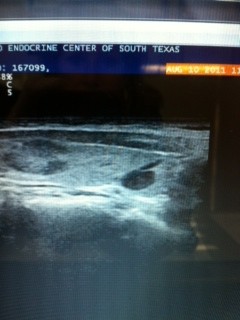
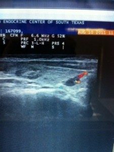 This picture is the right sagittal view on my 83 year old, otherwise healthy, woman with elevated calcium for several years when she can to me. Her elevated calcium was discovered when she broke her hip and was found to have osteoporosis from high calcium. Her sestimibi scan was negative, even her ultrasound was negative by her endocrinologist (not ECNU trained). She was followed conservatively for 3 years, till patient felt so bad she sought a second opinion. This picture clearly marks 2 nodules in her right thyroid lobe as well as an intra-thyroidal parathyroid with a polar vessel clearly feeding this lesion. This was not seen on sestimibi and mistaken for a thyroid nodule by her endocrinologist. She underwent a right thyroid lobectomy and her intra-operative PTH dropped from 123 to 26. Her irritability, depression and muscle cramps were resolved within 24 hours of surgery. Her bones will gain significant density over the next year, decreasing her risks for further fractures.

More Information
